Copyright
©The Author(s) 2017.
World J Gastroenterol. Jun 14, 2017; 23(22): 4127-4131
Published online Jun 14, 2017. doi: 10.3748/wjg.v23.i22.4127
Published online Jun 14, 2017. doi: 10.3748/wjg.v23.i22.4127
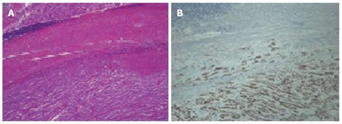
Figure 3 Histologic images.
A: The tumor in the mid-body consists of spindle cells. Above the spindle cell tumor, a smooth muscle layer, lymphoid cuffs, and fundic-type glands are found (× 40, HE); B: On immunohistochemical analysis, the spindle tumor cells are positive for S-100, in contrast to the overlying smooth muscles fibers and lymphoid cells (× 100).
- Citation: Choi KW, Joo M, Kim HS, Lee WY. Synchronous triple occurrence of MALT lymphoma, schwannoma, and adenocarcinoma of the stomach. World J Gastroenterol 2017; 23(22): 4127-4131
- URL: https://www.wjgnet.com/1007-9327/full/v23/i22/4127.htm
- DOI: https://dx.doi.org/10.3748/wjg.v23.i22.4127


