Copyright
©The Author(s) 2017.
World J Gastroenterol. Jun 14, 2017; 23(22): 4127-4131
Published online Jun 14, 2017. doi: 10.3748/wjg.v23.i22.4127
Published online Jun 14, 2017. doi: 10.3748/wjg.v23.i22.4127
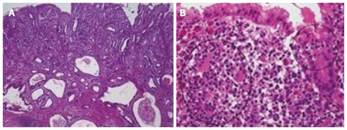
Figure 2 Histologic images.
A: The tumor in the antrum is a moderately differentiated adenocarcinoma [× 40, hematoxylin and eosin (HE)]; B: In the fundus section, lymphoepithelial lesions (circled in black), typical for mucosa-associated lymphoid tissue (MALT) lymphoma, are seen, which are formed by infiltration of centrocyte-like cells into the gastric glandular epithelium (× 400, HE).
- Citation: Choi KW, Joo M, Kim HS, Lee WY. Synchronous triple occurrence of MALT lymphoma, schwannoma, and adenocarcinoma of the stomach. World J Gastroenterol 2017; 23(22): 4127-4131
- URL: https://www.wjgnet.com/1007-9327/full/v23/i22/4127.htm
- DOI: https://dx.doi.org/10.3748/wjg.v23.i22.4127


