Copyright
©The Author(s) 2017.
World J Gastroenterol. Jun 14, 2017; 23(22): 4121-4126
Published online Jun 14, 2017. doi: 10.3748/wjg.v23.i22.4121
Published online Jun 14, 2017. doi: 10.3748/wjg.v23.i22.4121
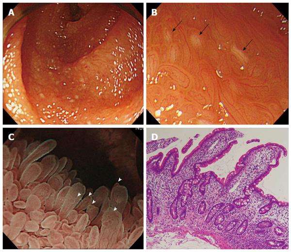
Figure 3 Trans-oral single balloon enteroscopy (jejunum) and histopathological findings.
A: White light observation demonstrated scattered white spots and reddening; B: Magnified observation under white light demonstrated scattered white spots (black arrows) as well as irregular caliber of the loop-like capillaries; C: Underwater magnifying enteroscopy demonstrated elongated villi and fine granular structures at the tips of villi (white arrowheads); D: Histopathological findings demonstrated twisted crypts and interstitial edema.
- Citation: Murata M, Bamba S, Takahashi K, Imaeda H, Nishida A, Inatomi O, Tsujikawa T, Kushima R, Sugimoto M, Andoh A. Application of novel magnified single balloon enteroscopy for a patient with Cronkhite-Canada syndrome. World J Gastroenterol 2017; 23(22): 4121-4126
- URL: https://www.wjgnet.com/1007-9327/full/v23/i22/4121.htm
- DOI: https://dx.doi.org/10.3748/wjg.v23.i22.4121


