Copyright
©The Author(s) 2017.
World J Gastroenterol. Jun 14, 2017; 23(22): 4121-4126
Published online Jun 14, 2017. doi: 10.3748/wjg.v23.i22.4121
Published online Jun 14, 2017. doi: 10.3748/wjg.v23.i22.4121
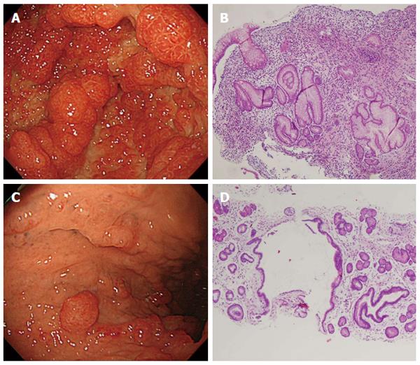
Figure 1 Esophagogastroduodenoscopy and histopathological findings.
A: Diffuse sessile and semipendunculated polyps were observed in the antrum; B: Biopsies of the polyps in the antrum indicated interstitial edematous and myxomatous lesions; C: Mild polyposis was observed in the upper body of the stomach; D: Biopsies of the interpolypoid lesions indicated the presence of the same interstitial edematous and myxomatous lesions as observed in polyp biopsies.
- Citation: Murata M, Bamba S, Takahashi K, Imaeda H, Nishida A, Inatomi O, Tsujikawa T, Kushima R, Sugimoto M, Andoh A. Application of novel magnified single balloon enteroscopy for a patient with Cronkhite-Canada syndrome. World J Gastroenterol 2017; 23(22): 4121-4126
- URL: https://www.wjgnet.com/1007-9327/full/v23/i22/4121.htm
- DOI: https://dx.doi.org/10.3748/wjg.v23.i22.4121


