Copyright
©The Author(s) 2017.
World J Gastroenterol. Jun 7, 2017; 23(21): 3928-3933
Published online Jun 7, 2017. doi: 10.3748/wjg.v23.i21.3928
Published online Jun 7, 2017. doi: 10.3748/wjg.v23.i21.3928
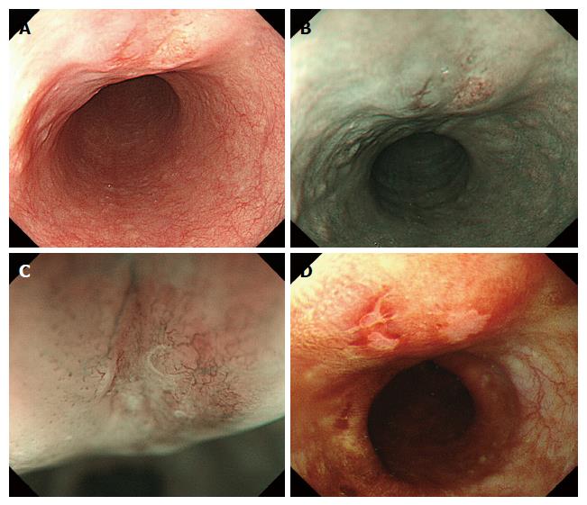
Figure 1 Endoscopic appearance.
A: White-light conventional endoscopy showed a submucosal tumor-like lesion. There were two reddish depressed areas on the surface of the tumor; B: The lesion appeared as brownish areas on NBI; C: Magnification endoscopy with NBI revealed irregular loop-shaped microvessels coexisting with irregularly branched thick non-looped vessels in depressed areas, where Lugol’s iodine (D) showed negative staining.
- Citation: Tamura H, Saiki H, Amano T, Yamamoto M, Hayashi S, Ando H, Doi R, Nishida T, Yamamoto K, Adachi S. Esophageal carcinoma originating in the surface epithelium with immunohistochemically proven esophageal gland duct differentiation: A case report. World J Gastroenterol 2017; 23(21): 3928-3933
- URL: https://www.wjgnet.com/1007-9327/full/v23/i21/3928.htm
- DOI: https://dx.doi.org/10.3748/wjg.v23.i21.3928


