Copyright
©The Author(s) 2017.
World J Gastroenterol. Jun 7, 2017; 23(21): 3825-3831
Published online Jun 7, 2017. doi: 10.3748/wjg.v23.i21.3825
Published online Jun 7, 2017. doi: 10.3748/wjg.v23.i21.3825
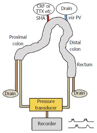
Figure 1 Schematic illustration of the isolated vascularly-perfused rat colon.
The isolated whole rat colon was placed in a temperature-controlled water bath and vascularly perfused with Krebs solution via the superior mesenteric artery. Luminal pressure was monitored via microtip catheter pressure transducers at the proximal and distal ends of the colon. Pressure changes were recorded with a data acquisition system. AT: Atropine sulfate; CRF: Corticotropin-releasing factor; PV: Portal vein; SMA: Superior mesenteric artery; TTX: Tetrodotoxin.
- Citation: Kim KJ, Kim KB, Yoon SM, Han JH, Chae HB, Park SM, Youn SJ. Corticotropin-releasing factor stimulates colonic motility via muscarinic receptors in the rat. World J Gastroenterol 2017; 23(21): 3825-3831
- URL: https://www.wjgnet.com/1007-9327/full/v23/i21/3825.htm
- DOI: https://dx.doi.org/10.3748/wjg.v23.i21.3825


