Copyright
©The Author(s) 2017.
World J Gastroenterol. May 28, 2017; 23(20): 3744-3751
Published online May 28, 2017. doi: 10.3748/wjg.v23.i20.3744
Published online May 28, 2017. doi: 10.3748/wjg.v23.i20.3744
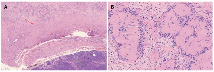
Figure 3 Microscopic examination.
A: A thin capsule (black arrow) was found between the tumor (red arrow) and the normal pancreatic tissues (green arrow) (HE, × 40); B: The tumor cells were spindle-shaped and had a palisading arrangement with no atypia, which is compatible with a benign tumor. Both hypercellular and hypocellular areas were visible (HE staining, × 200). HE: Hematoxylin and eosin.
- Citation: Xu SY, Wu YS, Li JH, Sun K, Hu ZH, Zheng SS, Wang WL. Successful treatment of a pancreatic schwannoma by spleen-preserving distal pancreatectomy. World J Gastroenterol 2017; 23(20): 3744-3751
- URL: https://www.wjgnet.com/1007-9327/full/v23/i20/3744.htm
- DOI: https://dx.doi.org/10.3748/wjg.v23.i20.3744


