Copyright
©The Author(s) 2017.
World J Gastroenterol. Jan 14, 2017; 23(2): 286-296
Published online Jan 14, 2017. doi: 10.3748/wjg.v23.i2.286
Published online Jan 14, 2017. doi: 10.3748/wjg.v23.i2.286
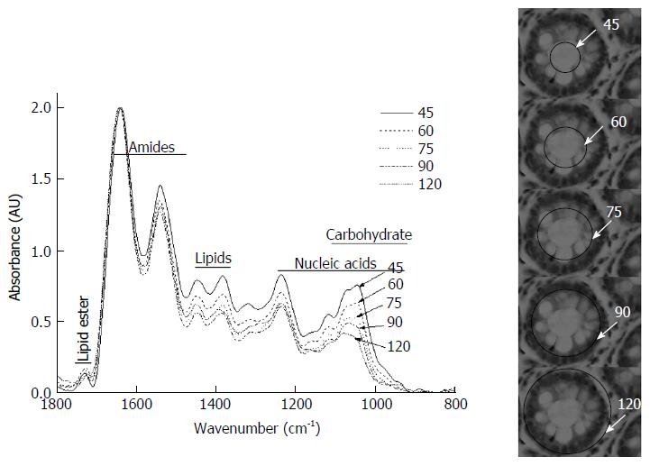
Figure 2 Amide I normalized spectra of a representative crypt with a lumen diameter of 45 μm and an outer diameter of 120 μm.
The histological sections on the right show the aperture of the microscope and the corresponding region of the crypt along with the spectra obtained for the same. The notations for the regions in the figure correspond to those of the spectra displayed in Figure 2. The spectra are normalized with respect to Amide I. Numbers indicate the size of the aperture.
- Citation: Sahu RK, Salman A, Mordechai S. Tracing overlapping biological signals in mid-infrared using colonic tissues as a model system. World J Gastroenterol 2017; 23(2): 286-296
- URL: https://www.wjgnet.com/1007-9327/full/v23/i2/286.htm
- DOI: https://dx.doi.org/10.3748/wjg.v23.i2.286


