Copyright
©The Author(s) 2017.
World J Gastroenterol. Jan 14, 2017; 23(2): 286-296
Published online Jan 14, 2017. doi: 10.3748/wjg.v23.i2.286
Published online Jan 14, 2017. doi: 10.3748/wjg.v23.i2.286
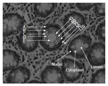
Figure 1 Histological section of H&E stained colonic biopsy showing the architecture of crypts in a transverse section.
The different zones indicated by letters show the part of the crypt that likely has a different metabolic pattern. The numbers indicate the size of the microscopic aperture that is used to obtain the spectra of the portion of the crypt. The circle consists of several cells joined in a concentric manner around the lumen with their nuclei in a peripheral configuration to the lumen. N: Nucleus; L: Lumen; C: Cytoplasm.
- Citation: Sahu RK, Salman A, Mordechai S. Tracing overlapping biological signals in mid-infrared using colonic tissues as a model system. World J Gastroenterol 2017; 23(2): 286-296
- URL: https://www.wjgnet.com/1007-9327/full/v23/i2/286.htm
- DOI: https://dx.doi.org/10.3748/wjg.v23.i2.286


