Copyright
©The Author(s) 2017.
World J Gastroenterol. Jan 14, 2017; 23(2): 275-285
Published online Jan 14, 2017. doi: 10.3748/wjg.v23.i2.275
Published online Jan 14, 2017. doi: 10.3748/wjg.v23.i2.275
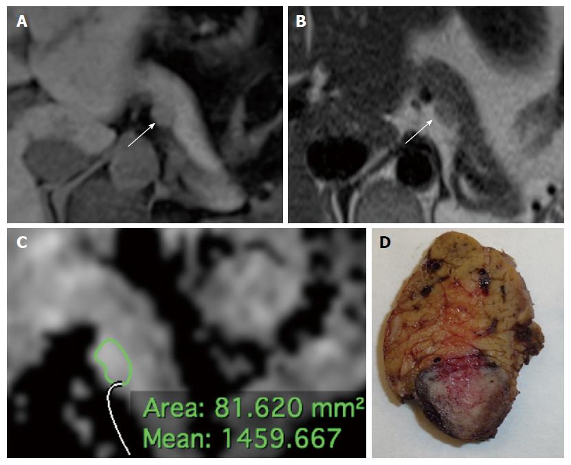
Figure 1 Small hyperfunctioning pancreatic neuroendocrine neoplasm (insulinoma) classified as G1, stage 1 tumor at histology (Ki67 < 1%, T1, N0, M0) in a 44 years-old woman.
A: Axial volumetric interpolated breath-hold examination (VIBE) gradient echo image (repetition time msec/echo time msec, 4.3/1.4) with fat suppression shows a homogeneously hypointense lesion with well defined margins (arrow) in the pancreatic body; B: On axial T2-weighted half-Fourier single-shot turbo spin echo (HASTE) image (TR/TE, ∞/90), the tumor appears slightly hyperintense (arrow) compared to adjacent pancreatic parenchyma; C: At ADC quantification the tumor had a relatively high mean ADC value; D: The macroscopic pathological specimen (distal pancreatectomy, sagittal cut) shows a small, well-delimitated lesion bulging the contour of the pancreatic body.
- Citation: De Robertis R, Cingarlini S, Tinazzi Martini P, Ortolani S, Butturini G, Landoni L, Regi P, Girelli R, Capelli P, Gobbo S, Tortora G, Scarpa A, Pederzoli P, D’Onofrio M. Pancreatic neuroendocrine neoplasms: Magnetic resonance imaging features according to grade and stage. World J Gastroenterol 2017; 23(2): 275-285
- URL: https://www.wjgnet.com/1007-9327/full/v23/i2/275.htm
- DOI: https://dx.doi.org/10.3748/wjg.v23.i2.275


