Copyright
©The Author(s) 2017.
World J Gastroenterol. May 21, 2017; 23(19): 3556-3564
Published online May 21, 2017. doi: 10.3748/wjg.v23.i19.3556
Published online May 21, 2017. doi: 10.3748/wjg.v23.i19.3556
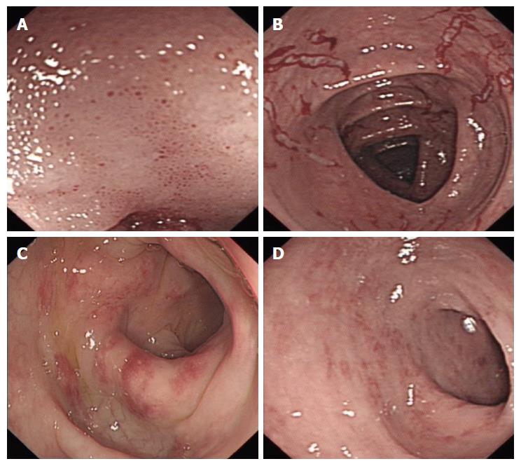
Figure 2 Endoscopic presentation of the eosinophilic gastroenteritis patients on colonoscopy.
A: Mucosal edema and small hemorrhagic spot in the distal ileum; B: Mucosal edema and erosions in the transverse colon; C: Segmental erythemoid edema and hyperemia in the descending colon; D: Erythemoid edema and hyperemia in the sigmoid colon.
- Citation: Abassa KK, Lin XY, Xuan JY, Zhou HX, Guo YW. Diagnosis of eosinophilic gastroenteritis is easily missed. World J Gastroenterol 2017; 23(19): 3556-3564
- URL: https://www.wjgnet.com/1007-9327/full/v23/i19/3556.htm
- DOI: https://dx.doi.org/10.3748/wjg.v23.i19.3556


