Copyright
©The Author(s) 2017.
World J Gastroenterol. May 14, 2017; 23(18): 3338-3348
Published online May 14, 2017. doi: 10.3748/wjg.v23.i18.3338
Published online May 14, 2017. doi: 10.3748/wjg.v23.i18.3338
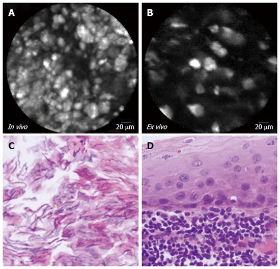
Figure 6 In vivo endoscopic ultrasound guided needle based confocal laser endomicroscopy, ex vivo confocal laser endomicroscopy, and histopathology of lymphoepithelial cyst.
Confocal laser endomicroscopy images, A (in vivo) and B (ex vivo) reveal clusters of bright particles representing keratin flakes. Macroscopically the lesion was filled with yellowish pasty material which by microscopy (panel C) demonstrated keratin flakes. The cyst was lined by squamous epithelium surrounded by abundant lymphoid tissue (panel D; HE, × 40).
- Citation: Krishna SG, Modi RM, Kamboj AK, Swanson BJ, Hart PA, Dillhoff ME, Manilchuk A, Schmidt CR, Conwell DL. In vivo and ex vivo confocal endomicroscopy of pancreatic cystic lesions: A prospective study. World J Gastroenterol 2017; 23(18): 3338-3348
- URL: https://www.wjgnet.com/1007-9327/full/v23/i18/3338.htm
- DOI: https://dx.doi.org/10.3748/wjg.v23.i18.3338


