Copyright
©The Author(s) 2017.
World J Gastroenterol. May 14, 2017; 23(18): 3338-3348
Published online May 14, 2017. doi: 10.3748/wjg.v23.i18.3338
Published online May 14, 2017. doi: 10.3748/wjg.v23.i18.3338
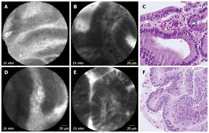
Figure 1 In vivo endoscopic ultrasound guided needle based confocal laser endomicroscopy, ex vivo confocal laser endomicroscopy, and histopathology of intraductal papillary mucinous neoplasms.
Panels A, B and C are from subject 1 (gastric subtype with high grade dysplasia). Panels D, E, and F are from subject 2 (intestinal subtype with high grade dysplasia). Complete “fingerlike” papillae are observed in both in vivo and ex vivo CLE. The vascular core in ex vivo CLE imaging is better defined. Histopathology (panels C, F): 40 × magnification; HE stain.
- Citation: Krishna SG, Modi RM, Kamboj AK, Swanson BJ, Hart PA, Dillhoff ME, Manilchuk A, Schmidt CR, Conwell DL. In vivo and ex vivo confocal endomicroscopy of pancreatic cystic lesions: A prospective study. World J Gastroenterol 2017; 23(18): 3338-3348
- URL: https://www.wjgnet.com/1007-9327/full/v23/i18/3338.htm
- DOI: https://dx.doi.org/10.3748/wjg.v23.i18.3338


