Copyright
©The Author(s) 2017.
World J Gastroenterol. May 7, 2017; 23(17): 3011-3016
Published online May 7, 2017. doi: 10.3748/wjg.v23.i17.3011
Published online May 7, 2017. doi: 10.3748/wjg.v23.i17.3011
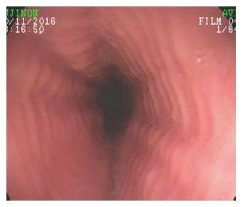
Figure 1 Eosinophilic esophagitis.
Endoscopic appearance of eosinophilic esophagitis; note the characteristic multiple rings throughout the esophagus resembling the tracheal aspect and described as “trachealization of the esophagus”. This finding is not common in the early stage of the disease, when tissue elasticity is still preserved by the inflammatory damage.
- Citation: Grossi L, Ciccaglione AF, Marzio L. Esophagitis and its causes: Who is “guilty” when acid is found “not guilty”? World J Gastroenterol 2017; 23(17): 3011-3016
- URL: https://www.wjgnet.com/1007-9327/full/v23/i17/3011.htm
- DOI: https://dx.doi.org/10.3748/wjg.v23.i17.3011


