Copyright
©The Author(s) 2017.
World J Gastroenterol. Apr 28, 2017; 23(16): 2899-2911
Published online Apr 28, 2017. doi: 10.3748/wjg.v23.i16.2899
Published online Apr 28, 2017. doi: 10.3748/wjg.v23.i16.2899
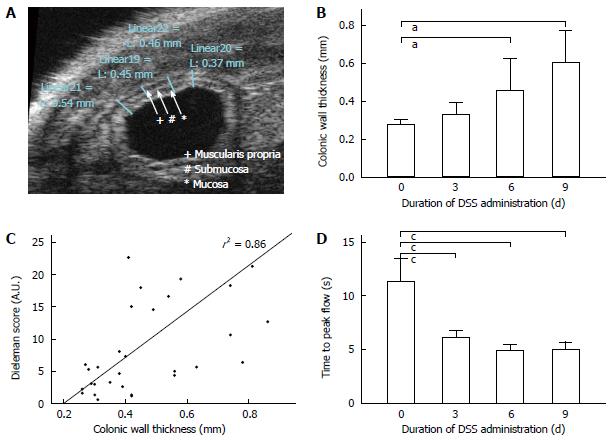
Figure 2 Contrast-enhanced ultrasound accurately detects dextran sodium-sulfate-induced colitis.
A: Native ultrasound of the murine colon visualizing the bowel wall stratification. Thickness of the wall was measured in triplicate. Colon filled with ultrasound contact gel for improved testing conditions; B: Colonic wall thicknesses correlate with exposure to DSS in a time-dependent manner. DSS treatment led to a loss of sonographic bowel wall stratification (not shown). Data are means ± SD, n =10 for each group, aP < 0.05; C: Linear regression between parameters thickness of colon wall and histological Dieleman score shows a significant correlation as indicated by determination coefficient r2 = 0.86; D: Time to peak flow depicting start of contrast agent application until maximal pixel contrast intensity in seconds. Time to peak flow correlates with exposure to DSS in a time-dependent manner. Data are mean ± SD, n = 10 for each group, cP < 0.01.
- Citation: Brückner M, Heidemann J, Nowacki TM, Cordes F, Stypmann J, Lenz P, Gohar F, Lügering A, Bettenworth D. Detection and characterization of murine colitis and carcinogenesis by molecularly targeted contrast-enhanced ultrasound. World J Gastroenterol 2017; 23(16): 2899-2911
- URL: https://www.wjgnet.com/1007-9327/full/v23/i16/2899.htm
- DOI: https://dx.doi.org/10.3748/wjg.v23.i16.2899


