Copyright
©The Author(s) 2017.
World J Gastroenterol. Apr 28, 2017; 23(16): 2899-2911
Published online Apr 28, 2017. doi: 10.3748/wjg.v23.i16.2899
Published online Apr 28, 2017. doi: 10.3748/wjg.v23.i16.2899
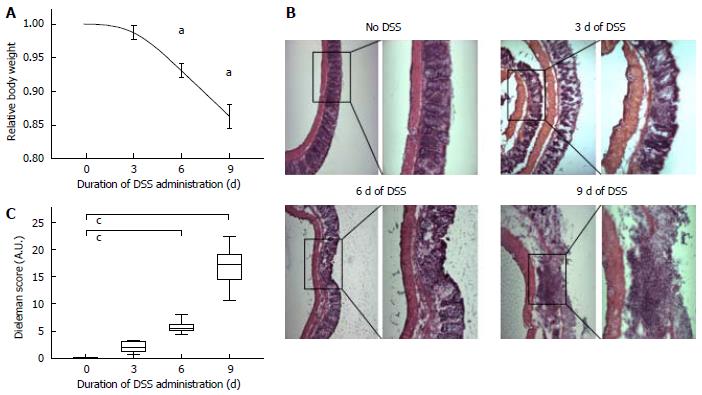
Figure 1 Assessment of dextran sodium-sulfate-incuded colitis by evaluation of relative body weight loss and histological damage.
C57BL/6 WT mice received 3% DSS in their drinking water for 3, 6 or 9 d, respectively. Inflammation was monitored by daily measurement of individual body weights. A: Development of relative body weight after 3, 6 and 9 d of treatment with DSS. Control group received tap water only. Data are mean ± SD; n = 10 for each group, aP < 0.01; B: Representative HE-stained histologic images of resected colons showing increasing inflammatory infiltrates (left panels, magnification × 10; right panels, magnification × 20); C: Histologic Dieleman Score (range from 0-40 points) describing inflammation, depth of inflammation and crypt damage or regeneration. Boxplot with median and whiskers, n = 10 for each group, cP < 0.01. DSS: Dextran sodium-sulfate.
- Citation: Brückner M, Heidemann J, Nowacki TM, Cordes F, Stypmann J, Lenz P, Gohar F, Lügering A, Bettenworth D. Detection and characterization of murine colitis and carcinogenesis by molecularly targeted contrast-enhanced ultrasound. World J Gastroenterol 2017; 23(16): 2899-2911
- URL: https://www.wjgnet.com/1007-9327/full/v23/i16/2899.htm
- DOI: https://dx.doi.org/10.3748/wjg.v23.i16.2899


