Copyright
©The Author(s) 2017.
World J Gastroenterol. Apr 21, 2017; 23(15): 2811-2818
Published online Apr 21, 2017. doi: 10.3748/wjg.v23.i15.2811
Published online Apr 21, 2017. doi: 10.3748/wjg.v23.i15.2811
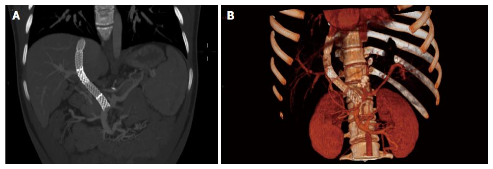
Figure 6 Computed tomography MIP multiplanar coronal reconstruction at 1 mo follow up (A and B).
A: Residual waist of the TIPS stent, regular portal liver perfusion and preservation of splenic and superior mesenteric venous flow. No residual free fluid in the perihepatic space; B: Volume rendering 3D computed tomography reconstruction: varice exclusion and global perspective of the regular portal flow.
- Citation: Pelizzo G, Quaretti P, Moramarco LP, Corti R, Maestri M, Iacob G, Calcaterra V. One step minilaparotomy-assisted transmesenteric portal vein recanalization combined with transjugular intrahepatic portosystemic shunt placement: A novel surgical proposal in pediatrics. World J Gastroenterol 2017; 23(15): 2811-2818
- URL: https://www.wjgnet.com/1007-9327/full/v23/i15/2811.htm
- DOI: https://dx.doi.org/10.3748/wjg.v23.i15.2811


