Copyright
©The Author(s) 2017.
World J Gastroenterol. Apr 21, 2017; 23(15): 2685-2695
Published online Apr 21, 2017. doi: 10.3748/wjg.v23.i15.2685
Published online Apr 21, 2017. doi: 10.3748/wjg.v23.i15.2685
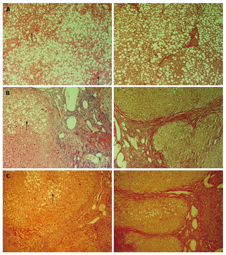
Figure 1 Histological staining of human liver tissue.
Representative images of donor tissue (A), NASH tissue (B) and ARLD liver (C) stained using haematoxylin and eosin (left panel) or Van Gieson stain (right panel). Bar = 100 μm and images were captured at 10 × original magnification. Data are representative of 6-14 samples in each group. Arrows in A indicate areas of localised inflammation present in our steatotic donor livers and arrows in B and C show steatotic hepatocytes. NASH: Nonalcoholic steatohepatitis; ARLD: Alcohol-related liver damage.
- Citation: Schofield Z, Reed MA, Newsome PN, Adams DH, Günther UL, Lalor PF. Changes in human hepatic metabolism in steatosis and cirrhosis. World J Gastroenterol 2017; 23(15): 2685-2695
- URL: https://www.wjgnet.com/1007-9327/full/v23/i15/2685.htm
- DOI: https://dx.doi.org/10.3748/wjg.v23.i15.2685


