Copyright
©The Author(s) 2017.
World J Gastroenterol. Apr 21, 2017; 23(15): 2673-2684
Published online Apr 21, 2017. doi: 10.3748/wjg.v23.i15.2673
Published online Apr 21, 2017. doi: 10.3748/wjg.v23.i15.2673
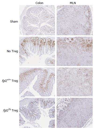
Figure 5 Fgl2Tg Treg prevent proliferation of infiltrating T cells in the colon.
CD3+ T cell proliferation in the MLN and colon were examined by Ki67 staining at week 14. Ki67+ cells were seen primarily in the cortex of the MLN of sham mice. Mice that received Teff had increased clusters of Ki67+ cells, primarily within the cortex. Mice that received fgl2+/+ Treg also had significant numbers of Ki67+ cells, whereas mice that received fgl2Tg Treg had similar numbers of Ki67+ cells as sham mice. Ki67+ cells were only seen in the colonic crypts of sham mice. Mice that were reconstituted with Teff alone had large numbers of Ki67+ cells both within the lamina propria and epithelium, coincident with areas of colonic inflammation. Mice that received fgl2+/+ Treg had small foci of Ki67+ cells in the lamina propria and epithelium whereas no Ki67+ staining was seen in these areas in mice that received fgl2Tg Treg. In all groups of mice colonic crypt cells stained positively for Ki67 as expected. Original magnification × 100. MLN: Mesenteric lymph nodes; Teff: Effector T cells; Treg: Regulatory T cells.
- Citation: Bartczak A, Zhang J, Adeyi O, Amir A, Grant D, Gorczynski R, Selzner N, Chruscinski A, Levy GA. Overexpression of fibrinogen-like protein 2 protects against T cell-induced colitis. World J Gastroenterol 2017; 23(15): 2673-2684
- URL: https://www.wjgnet.com/1007-9327/full/v23/i15/2673.htm
- DOI: https://dx.doi.org/10.3748/wjg.v23.i15.2673


