Copyright
©The Author(s) 2017.
World J Gastroenterol. Apr 14, 2017; 23(14): 2493-2504
Published online Apr 14, 2017. doi: 10.3748/wjg.v23.i14.2493
Published online Apr 14, 2017. doi: 10.3748/wjg.v23.i14.2493
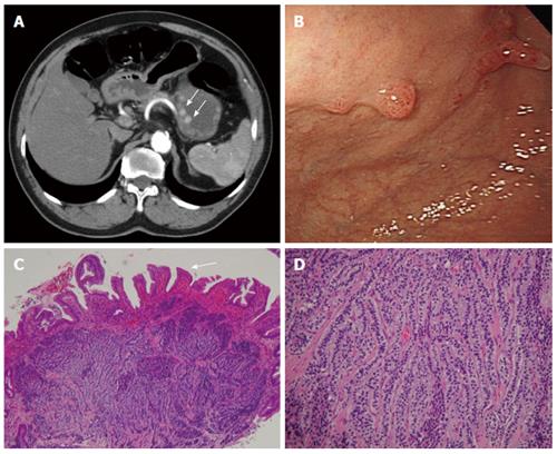
Figure 8 Carcinoid.
A 66-year-old man presented with epigastralgia and elevated levels of serum gastrin. A: Contrast-enhanced transverse and coronal computed tomography (CT) scans showing multiple enhancing polypoid lesions (arrows) at the gastric body; B: Endoscopy showing multiple polypoid lesions; C: Low-power photomicrograph (original magnification, × 10; HE stain) showing atrophic gastritis (atrophy in glandular structures, arrow); D: High-power photomicrograph (original magnification, × 100; HE stain) showing uniform cells bearing round nuclei and growing in a festoon or ribbon-like arrangement in the submucosa.
- Citation: Lin YM, Chiu NC, Li AFY, Liu CA, Chou YH, Chiou YY. Unusual gastric tumors and tumor-like lesions: Radiological with pathological correlation and literature review. World J Gastroenterol 2017; 23(14): 2493-2504
- URL: https://www.wjgnet.com/1007-9327/full/v23/i14/2493.htm
- DOI: https://dx.doi.org/10.3748/wjg.v23.i14.2493


