Copyright
©The Author(s) 2017.
World J Gastroenterol. Apr 14, 2017; 23(14): 2493-2504
Published online Apr 14, 2017. doi: 10.3748/wjg.v23.i14.2493
Published online Apr 14, 2017. doi: 10.3748/wjg.v23.i14.2493
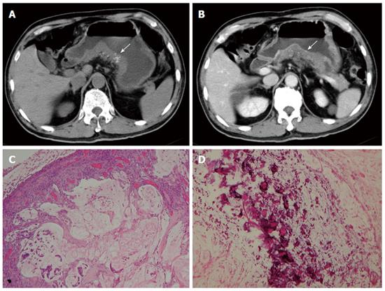
Figure 6 Mucinous adenocarcinoma.
A 65-year-old man presented with vomiting and diarrhea for 2 mo. A and B: Pre- and post-contrast-enhanced computed tomography scans shoings a segmental thickening at the posterior wall of the gastric antrum, with poor enhancement and punctate calcification (arrow). Low-power photomicrograph (original magnification, × 20; HE stain) showing abundant extracellular mucin pools (C) with floating tumor cells and calcifications (D).
- Citation: Lin YM, Chiu NC, Li AFY, Liu CA, Chou YH, Chiou YY. Unusual gastric tumors and tumor-like lesions: Radiological with pathological correlation and literature review. World J Gastroenterol 2017; 23(14): 2493-2504
- URL: https://www.wjgnet.com/1007-9327/full/v23/i14/2493.htm
- DOI: https://dx.doi.org/10.3748/wjg.v23.i14.2493


