Copyright
©The Author(s) 2017.
World J Gastroenterol. Apr 14, 2017; 23(14): 2493-2504
Published online Apr 14, 2017. doi: 10.3748/wjg.v23.i14.2493
Published online Apr 14, 2017. doi: 10.3748/wjg.v23.i14.2493
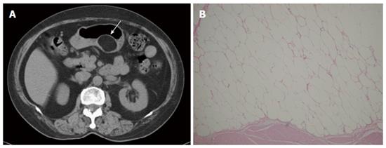
Figure 4 Lipoma.
A 69-year-old man presented with abdominal fullness. A: Non-contrast-enhanced computed tomography (CT) showing a round, sharply marginated, uniform fatty mass (arrow) with negative CT numbers (-90 HU) in the greater curvature of the stomach; B: High-power photomicrograph (original magnification, × 200; HE stain) showing that the tumor consists of mature adipocytes.
- Citation: Lin YM, Chiu NC, Li AFY, Liu CA, Chou YH, Chiou YY. Unusual gastric tumors and tumor-like lesions: Radiological with pathological correlation and literature review. World J Gastroenterol 2017; 23(14): 2493-2504
- URL: https://www.wjgnet.com/1007-9327/full/v23/i14/2493.htm
- DOI: https://dx.doi.org/10.3748/wjg.v23.i14.2493


