Copyright
©The Author(s) 2017.
World J Gastroenterol. Apr 14, 2017; 23(14): 2493-2504
Published online Apr 14, 2017. doi: 10.3748/wjg.v23.i14.2493
Published online Apr 14, 2017. doi: 10.3748/wjg.v23.i14.2493
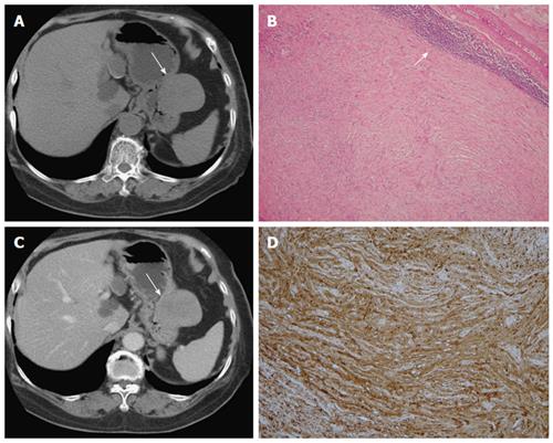
Figure 2 Schwannoma.
A 75-year-old woman presented with coffee ground vomitus. A: Pre-contrast transverse computed tomography (CT) showing a homogeneous iso-density tumor in the greater curvature of the stomach (arrow); B: Post-contrast-enhanced CT showing homogeneously moderate enhancement with a mixed (endoluminal and exophytic) growth pattern; C: Low-power photomicrograph (original magnification, × 20; HE stain) showing that the tumor retains its circumscription with lymphoid aggregate cuffing (arrow); D: The vaguely bundled spindle tumor cells were positive for S-100.
- Citation: Lin YM, Chiu NC, Li AFY, Liu CA, Chou YH, Chiou YY. Unusual gastric tumors and tumor-like lesions: Radiological with pathological correlation and literature review. World J Gastroenterol 2017; 23(14): 2493-2504
- URL: https://www.wjgnet.com/1007-9327/full/v23/i14/2493.htm
- DOI: https://dx.doi.org/10.3748/wjg.v23.i14.2493


