Copyright
©The Author(s) 2017.
World J Gastroenterol. Apr 7, 2017; 23(13): 2443-2447
Published online Apr 7, 2017. doi: 10.3748/wjg.v23.i13.2443
Published online Apr 7, 2017. doi: 10.3748/wjg.v23.i13.2443
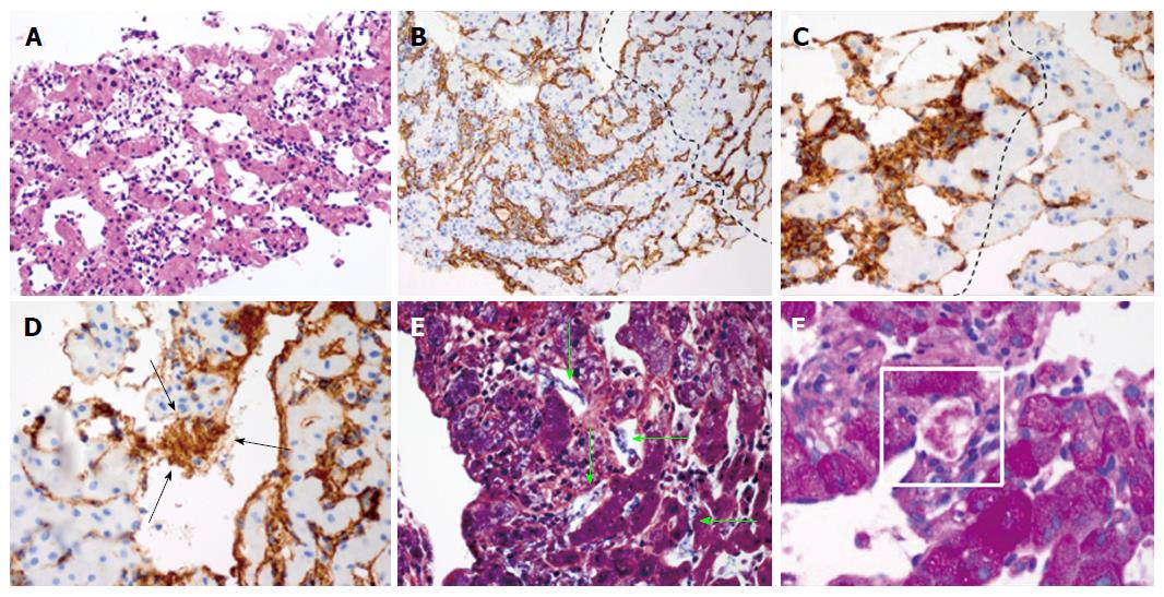
Figure 4 Histopathological findings of tumor needle biopsy suggest the presence of Kasabach-Merritt syndrome.
A: Hematoxylin-eosin (H&E) staining of tumor demonstrates dilated vascular channels lined by atypical endothelial cells with hyperchromatic, enlarged nuclei and reticular cytoplasm; B: Immunohistochemical stain for vascular antigen CD34 showing diffuse infiltration of CD34+ cells throughout the sinusoids in the tumor (left of line) with focal aggregation. Uninvolved region (right of line) shows normal liver sinusoidal endothelial cells (LSEC) that highlight the nondilated sinusoids along the cord of hepatocytes; C and D: Immunohistochemical stain of von-Willebrand Factor (vWF)/Factor VIII shows increased expression within tumor cells (left of line) as compared to uninvolved region (right of line). Extracellular aggregate positive for vWF/Factor VIII is seen within dilated sinusoid of angiosarcoma (arrow); E: Phophotungistic acid-hematoxylin stain (PTAH) demonstrates fibrin nets within the tumor seen as extracellular fibrillary structures that stain blue (arrow); F: Periodic acid-Schiff (PAS) stain of the tumor demonstrates glycogen granules within extracellular material of vascular channels, representing clumps of entrapped platelets (shown in rectangle). Note that positive PAS staining of glycogen is also observed in native hepatocytes.
- Citation: Wadhwa S, Kim TH, Lin L, Kanel G, Saito T. Hepatic angiosarcoma with clinical and histological features of Kasabach-Merritt syndrome. World J Gastroenterol 2017; 23(13): 2443-2447
- URL: https://www.wjgnet.com/1007-9327/full/v23/i13/2443.htm
- DOI: https://dx.doi.org/10.3748/wjg.v23.i13.2443


