Copyright
©The Author(s) 2017.
World J Gastroenterol. Apr 7, 2017; 23(13): 2443-2447
Published online Apr 7, 2017. doi: 10.3748/wjg.v23.i13.2443
Published online Apr 7, 2017. doi: 10.3748/wjg.v23.i13.2443
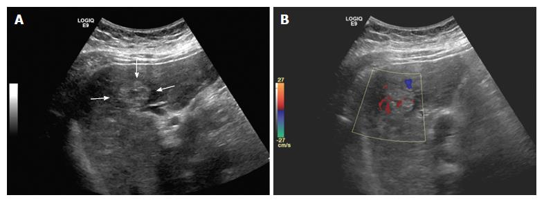
Figure 1 Ultrasonographic findings of hepatic angiosarcoma.
A: Representative image of tumor detected by abdominal ultrasound (arrow). Note the increased peripheral echogenicity and hypoechoic central appearance; B: Color doppler study of (A) demonstrates hypervascularity of the tumor. No arterial flow was detected (image not shown).
- Citation: Wadhwa S, Kim TH, Lin L, Kanel G, Saito T. Hepatic angiosarcoma with clinical and histological features of Kasabach-Merritt syndrome. World J Gastroenterol 2017; 23(13): 2443-2447
- URL: https://www.wjgnet.com/1007-9327/full/v23/i13/2443.htm
- DOI: https://dx.doi.org/10.3748/wjg.v23.i13.2443


