Copyright
©The Author(s) 2017.
World J Gastroenterol. Apr 7, 2017; 23(13): 2414-2423
Published online Apr 7, 2017. doi: 10.3748/wjg.v23.i13.2414
Published online Apr 7, 2017. doi: 10.3748/wjg.v23.i13.2414
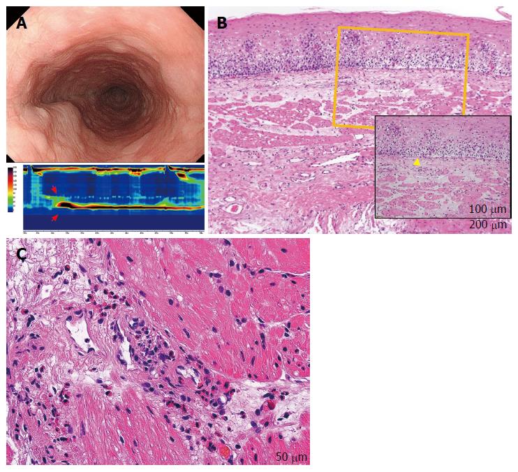
Figure 4 Eosinophilic esophageal myositis (case 10).
A: Endoscopy showing a compressed lumen similar to that in a case of subepithelial eosinophilic esophagitis (sEoE) and different to that in a case of eosinophilic esophagitis (EoE). Results of the high-resolution manometry are shown in the lower insert. An extremely high maximum distal contractile integral (DCI) is observable (13462.2 mmHg/[s•cm], red arrows). The patient was diagnosed with a jackhammer esophagus based on the Chicago classification criteria; B: The entry site for peroral esophageal muscle biopsy (POEM-b) was created by cap-fitted endoscopic mucosal resection in order to obtain mucosal and submucosal specimens. Epithelial eosinophilia was not seen, with only basal layer destruction (yellow triangle) apparent. In the subepithelial layer, the number of eosinophils was elevated, although the eosinophil number was less than that in the muscle layer; C: Muscle-layer histopathology by POEM-b, showing a severe eosinophilic infiltration in the perivascular and perimysial area of the muscle specimens (48 eosinophils per high-power field) at 400 × magnification.
- Citation: Sato H, Nakajima N, Takahashi K, Hasegawa G, Mizuno KI, Hashimoto S, Ikarashi S, Hayashi K, Honda Y, Yokoyama J, Sato Y, Terai S. Proposed criteria to differentiate heterogeneous eosinophilic gastrointestinal disorders of the esophagus, including eosinophilic esophageal myositis. World J Gastroenterol 2017; 23(13): 2414-2423
- URL: https://www.wjgnet.com/1007-9327/full/v23/i13/2414.htm
- DOI: https://dx.doi.org/10.3748/wjg.v23.i13.2414


