Copyright
©The Author(s) 2017.
World J Gastroenterol. Apr 7, 2017; 23(13): 2414-2423
Published online Apr 7, 2017. doi: 10.3748/wjg.v23.i13.2414
Published online Apr 7, 2017. doi: 10.3748/wjg.v23.i13.2414
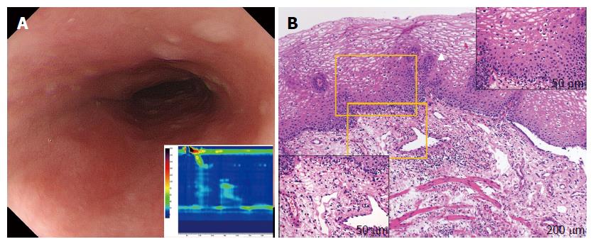
Figure 3 Subepithelial eosinophilic esophagitis (case 6).
A: Endoscopy showing the compressed lumen. Characteristic findings for eosinophilic esophagitis such as longitudinal furrows, white plaques, or fixed rings are not seen. High-resolution manometry (HRM) results, showing highly disrupted distal contractile integral (42.5 mmHg/[s•cm], lower insert). The patient was diagnosed with failed peristalsis based on the Chicago classification criteria; B: Mucosal histology by conventional biopsy at 100 × magnification. The white triangle indicates upward papillae elongation. In the upper-right inset, there is an absence of eosinophil infiltration in the epithelium (400 × magnification). Subepithelial eosinophilia (35 eosinophils per high-power field) is shown in the left lower inset (400 × magnification).
- Citation: Sato H, Nakajima N, Takahashi K, Hasegawa G, Mizuno KI, Hashimoto S, Ikarashi S, Hayashi K, Honda Y, Yokoyama J, Sato Y, Terai S. Proposed criteria to differentiate heterogeneous eosinophilic gastrointestinal disorders of the esophagus, including eosinophilic esophageal myositis. World J Gastroenterol 2017; 23(13): 2414-2423
- URL: https://www.wjgnet.com/1007-9327/full/v23/i13/2414.htm
- DOI: https://dx.doi.org/10.3748/wjg.v23.i13.2414


