Copyright
©The Author(s) 2017.
World J Gastroenterol. Apr 7, 2017; 23(13): 2414-2423
Published online Apr 7, 2017. doi: 10.3748/wjg.v23.i13.2414
Published online Apr 7, 2017. doi: 10.3748/wjg.v23.i13.2414
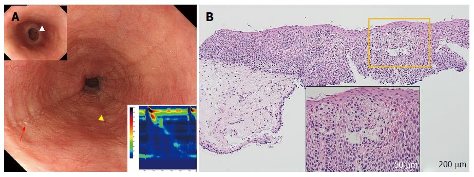
Figure 2 Eosinophilic esophagitis (case 3).
A: Endoscopy shows longitudinal furrows (yellow triangles) and white plaques (red arrow) in the medial esophagus, and the presence of fixed rings in the lower esophagus [upper insert (white triangle)]. High-resolution manometry (HRM) results showing highly disrupted distal contractile integral (DCI; 54.8 mmHg/[s•cm], lower insert). The patient was diagnosed with failed peristalsis based on the Chicago classification criteria; B: Histology at 100 × magnification, showing significant eosinophil inflammation was observed (50 eosinophils per high-power field) in the esophageal epithelium with dilated intercellular spaces (lower insert).
- Citation: Sato H, Nakajima N, Takahashi K, Hasegawa G, Mizuno KI, Hashimoto S, Ikarashi S, Hayashi K, Honda Y, Yokoyama J, Sato Y, Terai S. Proposed criteria to differentiate heterogeneous eosinophilic gastrointestinal disorders of the esophagus, including eosinophilic esophageal myositis. World J Gastroenterol 2017; 23(13): 2414-2423
- URL: https://www.wjgnet.com/1007-9327/full/v23/i13/2414.htm
- DOI: https://dx.doi.org/10.3748/wjg.v23.i13.2414


