Copyright
©The Author(s) 2017.
World J Gastroenterol. Apr 7, 2017; 23(13): 2318-2329
Published online Apr 7, 2017. doi: 10.3748/wjg.v23.i13.2318
Published online Apr 7, 2017. doi: 10.3748/wjg.v23.i13.2318
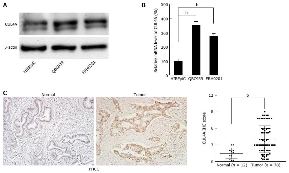
Figure 1 Cullin 4A is overexpressed in perihilar cholangiocarcinoma.
A: Expression of CUL4A protein was detected in normal biliary epithelial cells (HIBEpic) and PHCC cell lines by Western blot assays; B: Expression of CUL4A mRNA was detected in normal biliary epithelial cells (HIBEpiC) and PHCC cell lines by qRT-PCR assay; C: Representative images of CUL4A IHC staining in PHCC tumour tissues and normal adjacent tissues. Corresponding semiquantification of CUL4A expression is shown. Numbers in (B) indicate the fold changes of band densities based on at least three independent experiments. bP < 0.01 based on the Student’s t-test. Data are represented as mean ± SD. CUL4A: Cullin 4A; PHCC: Perihilar cholangiocarcinoma.
- Citation: Zhang TJ, Xue D, Zhang CD, Zhang ZD, Liu QR, Wang JQ. Cullin 4A is associated with epithelial to mesenchymal transition and poor prognosis in perihilar cholangiocarcinoma. World J Gastroenterol 2017; 23(13): 2318-2329
- URL: https://www.wjgnet.com/1007-9327/full/v23/i13/2318.htm
- DOI: https://dx.doi.org/10.3748/wjg.v23.i13.2318


