Copyright
©The Author(s) 2017.
World J Gastroenterol. Mar 28, 2017; 23(12): 2251-2257
Published online Mar 28, 2017. doi: 10.3748/wjg.v23.i12.2251
Published online Mar 28, 2017. doi: 10.3748/wjg.v23.i12.2251
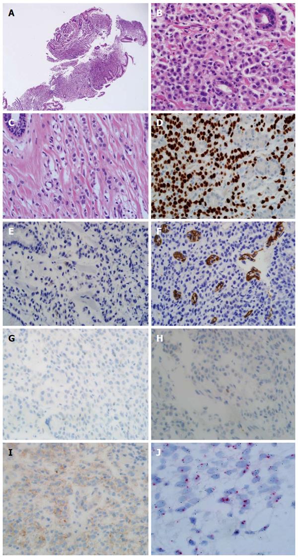
Figure 3 Pathologic features of endoscopic biopsy specimen.
Discohesive tumor cells are infiltrated in the stroma of the stomach mucosal tissue (HE × 40, A). Tumor cells show enlarged centrally located nucleus without intracytoplasmic clear mucin. The tumor cells had no connection to the remained normal gastric mucosal tissue (HE × 400, B). Previous breast cancer pathology was reviewed (C). Discohesive tumor cells were arranged in indian file. The tumor cells had enlarged centrally located nucleus without intracytoplasmic mucin (HE × 400, C). Immunohistochemical stains and molecular test of tumor was done (D-J). Diffuse strong nucleus expression of GATA3 was observed (GATA3 × 400, D). Focal, less than one percentage cytoplasmic expression of GCDFP was detected (GCDFP × 400, E). Negative stain for E-cadherin (E-cadherin × 400, F). Negative stains for ER and PR (ER × 400, PR × 400, G, H). Immunohistochemical stain for HER-2 was equivocal (HER-2 × 400, I). Silver in situ hybridization (SISH) for determination of HER2 gene status. Occasional HER2 gene amplified cells were noted in the mixture with normal HE2 gene expressing cells (SISH × 1000, J).
- Citation: Yim K, Ro SM, Lee J. Breast cancer metastasizing to the stomach mimicking primary gastric cancer: A case report. World J Gastroenterol 2017; 23(12): 2251-2257
- URL: https://www.wjgnet.com/1007-9327/full/v23/i12/2251.htm
- DOI: https://dx.doi.org/10.3748/wjg.v23.i12.2251


