Copyright
©The Author(s) 2017.
World J Gastroenterol. Mar 14, 2017; 23(10): 1920-1924
Published online Mar 14, 2017. doi: 10.3748/wjg.v23.i10.1920
Published online Mar 14, 2017. doi: 10.3748/wjg.v23.i10.1920
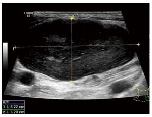
Figure 2 Ultrasonography of cervical mass: The mass was hypoechoic, with a smooth border and un-even internal echo.
- Citation: Ma C, Hao SL, Liu XC, Nin JY, Wu GC, Jiang LX, Fancellu A, Porcu A, Zheng HT. Supraclavicular lymph node metastases from malignant gastrointestinal stromal tumor of the jejunum: A case report with review of the literature. World J Gastroenterol 2017; 23(10): 1920-1924
- URL: https://www.wjgnet.com/1007-9327/full/v23/i10/1920.htm
- DOI: https://dx.doi.org/10.3748/wjg.v23.i10.1920


