Copyright
©The Author(s) 2017.
World J Gastroenterol. Mar 14, 2017; 23(10): 1735-1746
Published online Mar 14, 2017. doi: 10.3748/wjg.v23.i10.1735
Published online Mar 14, 2017. doi: 10.3748/wjg.v23.i10.1735
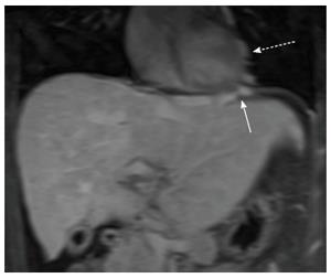
Figure 6 Coronal post-gadolinium T1-weighted fat-suppressed magnetic resonance image shows prominent cardiophrenic (white arrow) and pericardial collateral veins (dashed white arrow) in a patient with Budd-Chiari syndrome.
- Citation: Bandali MF, Mirakhur A, Lee EW, Ferris MC, Sadler DJ, Gray RR, Wong JK. Portal hypertension: Imaging of portosystemic collateral pathways and associated image-guided therapy. World J Gastroenterol 2017; 23(10): 1735-1746
- URL: https://www.wjgnet.com/1007-9327/full/v23/i10/1735.htm
- DOI: https://dx.doi.org/10.3748/wjg.v23.i10.1735


