Copyright
©The Author(s) 2017.
World J Gastroenterol. Mar 14, 2017; 23(10): 1735-1746
Published online Mar 14, 2017. doi: 10.3748/wjg.v23.i10.1735
Published online Mar 14, 2017. doi: 10.3748/wjg.v23.i10.1735
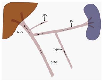
Figure 1 Normal portal venous anatomy and direction of blood flow.
The main portal vein (MPV) is most commonly formed when the splenic vein (SV) and the superior mesenteric vein (SMV) join. While variable, the inferior mesenteric vein (IMV) most commonly drains in to the splenic vein, at the level of the pancreatic body. Other tributaries may also join the MPV, such as the left gastric vein (LGV) as depicted here.
- Citation: Bandali MF, Mirakhur A, Lee EW, Ferris MC, Sadler DJ, Gray RR, Wong JK. Portal hypertension: Imaging of portosystemic collateral pathways and associated image-guided therapy. World J Gastroenterol 2017; 23(10): 1735-1746
- URL: https://www.wjgnet.com/1007-9327/full/v23/i10/1735.htm
- DOI: https://dx.doi.org/10.3748/wjg.v23.i10.1735


