Copyright
©The Author(s) 2017.
World J Gastroenterol. Jan 7, 2017; 23(1): 25-41
Published online Jan 7, 2017. doi: 10.3748/wjg.v23.i1.25
Published online Jan 7, 2017. doi: 10.3748/wjg.v23.i1.25
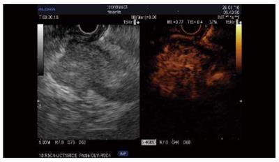
Figure 2 Contrast-enhanced harmonics-endoscopic ultrasound of a neuroendocrine pancreatic tumor.
Standard endoscopic ultrasound image (left) of a hypoenhanced, well-delineated lesion of the head of the pancreas. The contrast image (right) shows a hyperenhanced lesion that is suggestive of a neuroendocrine tumor, as later proved by fine needle aspiration.
- Citation: Seicean A, Mosteanu O, Seicean R. Maximizing the endosonography: The role of contrast harmonics, elastography and confocal endomicroscopy. World J Gastroenterol 2017; 23(1): 25-41
- URL: https://www.wjgnet.com/1007-9327/full/v23/i1/25.htm
- DOI: https://dx.doi.org/10.3748/wjg.v23.i1.25


