Copyright
©The Author(s) 2016.
World J Gastroenterol. Mar 7, 2016; 22(9): 2855-2860
Published online Mar 7, 2016. doi: 10.3748/wjg.v22.i9.2855
Published online Mar 7, 2016. doi: 10.3748/wjg.v22.i9.2855
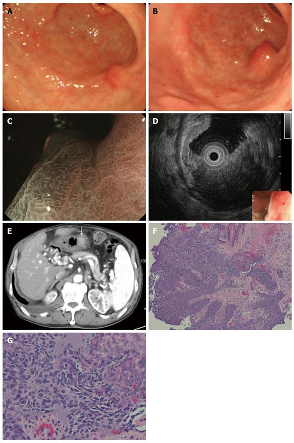
Figure 4 Follow-up esophagogastroduodenoscopy after endoscopic submucosal dissection.
A: A follow-up esophagogastroduodenoscopy (EGD) 7 mo after endoscopic submucosal dissection (ESD) showed a slight elevation on the post ESD scar, but biopsy specimens did not show any malignancy; B: Two more months later, EGD showed a submucosal tumor-like lesion with ulceration 15 mm in size, located right under the post ESD scar; C: Magnifying endoscopy with narrow band imaging showed that the base of the elevation was covered with normal mucosa; D: EUS findings indicated that the tumor mainly involved the submucosal layer; E: The tumor was also recognized as well-demarcated, early enhanced mass on CT; F, G: Biopsy specimens from the edge of the ulcer on the top of the elevated lesion revealed moderately differentiated SCC, which resembled the EC (magnification: F × 40, G × 200).
- Citation: Asai S, Takeshita K, Kano Y, Nakao E, Ichinona T, Fujimoto N, Akamine E, Mori T, Ogawa A. Implantation of esophageal cancer onto post-dissection ulcer after gastric endoscopic submucosal dissection. World J Gastroenterol 2016; 22(9): 2855-2860
- URL: https://www.wjgnet.com/1007-9327/full/v22/i9/2855.htm
- DOI: https://dx.doi.org/10.3748/wjg.v22.i9.2855


