Copyright
©The Author(s) 2016.
World J Gastroenterol. Mar 7, 2016; 22(9): 2855-2860
Published online Mar 7, 2016. doi: 10.3748/wjg.v22.i9.2855
Published online Mar 7, 2016. doi: 10.3748/wjg.v22.i9.2855
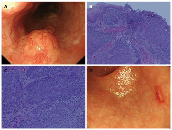
Figure 1 Esophagogastroduodenoscopy findings.
A: An elevated lesion 20 mm in size was detected in the left wall of the middle thoracic esophagus. The endoscopic findings indicated that the lesion was esophageal cancer with a depth of SM2; B, C: Biopsy specimens revealed moderately differentiated squamous cell carcinoma (hematoxylin-eosin stain; magnification: B × 40, C × 200); D: A small erosion with surrounding reddish mucosa was also found in the posterior wall of the gastric antrum, which was suspected to be gastric mucosal cancer.
- Citation: Asai S, Takeshita K, Kano Y, Nakao E, Ichinona T, Fujimoto N, Akamine E, Mori T, Ogawa A. Implantation of esophageal cancer onto post-dissection ulcer after gastric endoscopic submucosal dissection. World J Gastroenterol 2016; 22(9): 2855-2860
- URL: https://www.wjgnet.com/1007-9327/full/v22/i9/2855.htm
- DOI: https://dx.doi.org/10.3748/wjg.v22.i9.2855


