Copyright
©The Author(s) 2016.
World J Gastroenterol. Mar 7, 2016; 22(9): 2760-2770
Published online Mar 7, 2016. doi: 10.3748/wjg.v22.i9.2760
Published online Mar 7, 2016. doi: 10.3748/wjg.v22.i9.2760
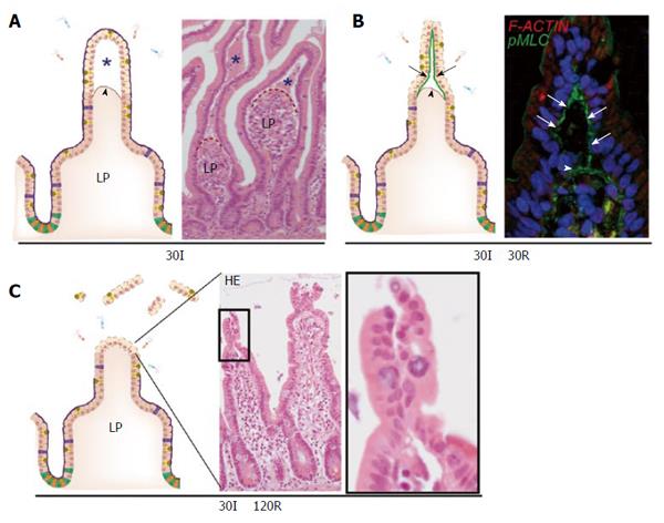
Figure 2 The human small intestine has efficient mechanisms to prevent excessive epithelial lining damage.
A: Cartoon and hematoxylin-eosin staining demonstrating the appearance of subepithelial spaces (asterisks) after ischemia as a result of retraction of the basement membrane (arrowhead); B: Early during reperfusion, loose IR-damaged epithelial sheets are pulled together through active contraction of pMLC at the basal side of epithelial cells (green line, arrows), bringing these cells together; C: Zipper-like constriction of the epithelium is associated with rapid restoration of the epithelial lining and prevents exposure of lamina propria to intraluminal content. LP: Lamina propria; pMLC: Phosphorylated myosin light chain; F-ACTIN: Filamentous actin.
- Citation: Grootjans J, Lenaerts K, Buurman WA, Dejong CHC, Derikx JPM. Life and death at the mucosal-luminal interface: New perspectives on human intestinal ischemia-reperfusion. World J Gastroenterol 2016; 22(9): 2760-2770
- URL: https://www.wjgnet.com/1007-9327/full/v22/i9/2760.htm
- DOI: https://dx.doi.org/10.3748/wjg.v22.i9.2760


