Copyright
©The Author(s) 2016.
World J Gastroenterol. Mar 7, 2016; 22(9): 2678-2700
Published online Mar 7, 2016. doi: 10.3748/wjg.v22.i9.2678
Published online Mar 7, 2016. doi: 10.3748/wjg.v22.i9.2678
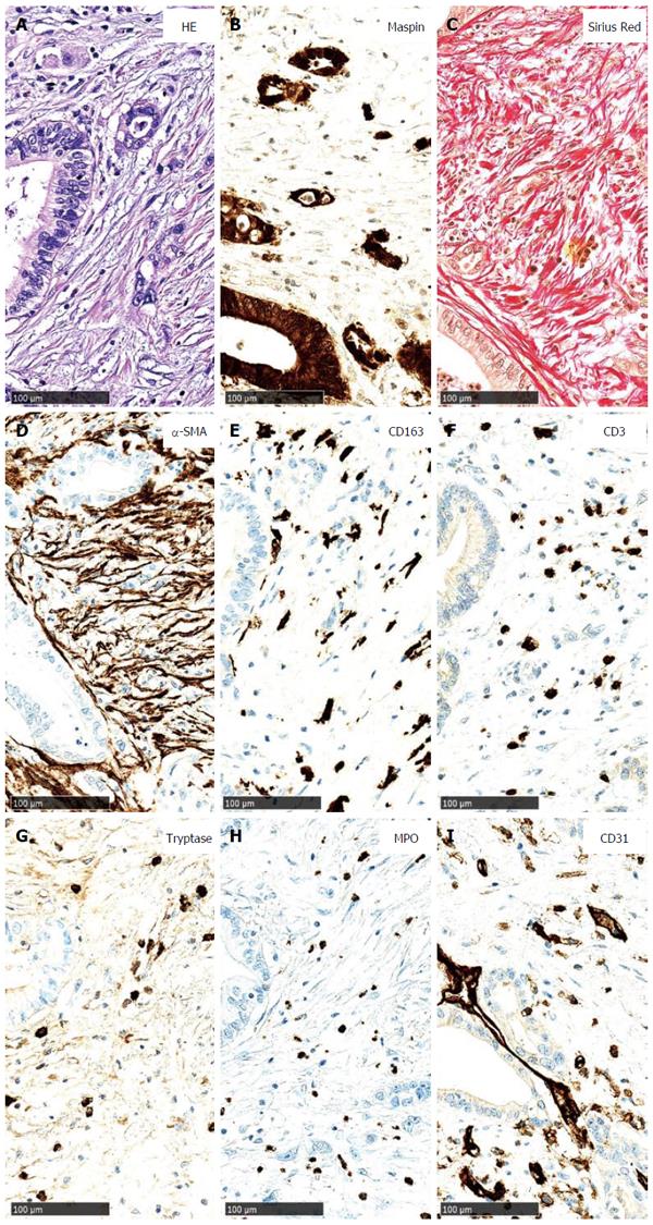
Figure 4 Main components of the tumour stroma in pancreatic cancer.
A: Pancreatic cancer (PC) cells, arranged in small groups and duct-like structures, are surrounded by desmoplastic stroma (HE staining); B: PC cells strongly express maspin (maspin immunostaining); C: Using a Sirius red stain, the collagen fibres of the desmoplastic stroma are highlighted; D: Numerous α-smooth muscle actin-positive cancer-associated fibroblasts are observed; E: Tumour-infiltrating macrophages (CD163 immunostaining); F: T cells (CD3 immunostaining) are shown; G: Additionally, a few mast cells (tryptase immunostaining); H: neutrophilic granulocytes (myeloperoxidase immunostaining) are present; I: Several newly formed small blood vessels are located in the desmoplastic stroma (CD31 immunostaining).
- Citation: Nielsen MFB, Mortensen MB, Detlefsen S. Key players in pancreatic cancer-stroma interaction: Cancer-associated fibroblasts, endothelial and inflammatory cells. World J Gastroenterol 2016; 22(9): 2678-2700
- URL: https://www.wjgnet.com/1007-9327/full/v22/i9/2678.htm
- DOI: https://dx.doi.org/10.3748/wjg.v22.i9.2678


