Copyright
©The Author(s) 2016.
World J Gastroenterol. Feb 28, 2016; 22(8): 2636-2641
Published online Feb 28, 2016. doi: 10.3748/wjg.v22.i8.2636
Published online Feb 28, 2016. doi: 10.3748/wjg.v22.i8.2636
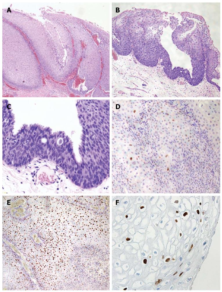
Figure 3 Histologic and immunohistochemical analyses of a specimen from the anal canal.
A: A low-power scan of the glass slide showing papillomatosis, parakeratosis, dyskeratosis, acanthosis, and distinct koilocytosis; B: A low-power scan shows an underlying chronic inflammatory cell infiltrate and basal cell atypical hyperplasia; C: Condylomata acuminatum with accentuated transepithelial lymphocytic infiltrate was diagnosed as anal intraepithelial neoplasia 2; D: Immunohistochemical peroxidase staining for p53 protein showing negative cells; E: Ki-67 stain showing 40% positive cells through the layer of the epithelium; F: Immunohistochemical studies indicate positive staining for papillomavirus common antigen.
- Citation: Sasaki A, Nakajima T, Egashira H, Takeda K, Tokoro S, Ichita C, Masuda S, Uojima H, Koizumi K, Kinbara T, Sakamoto T, Saito Y, Kako M. Condyloma acuminatum of the anal canal, treated with endoscopic submucosal dissection. World J Gastroenterol 2016; 22(8): 2636-2641
- URL: https://www.wjgnet.com/1007-9327/full/v22/i8/2636.htm
- DOI: https://dx.doi.org/10.3748/wjg.v22.i8.2636


