Copyright
©The Author(s) 2016.
World J Gastroenterol. Feb 28, 2016; 22(8): 2545-2557
Published online Feb 28, 2016. doi: 10.3748/wjg.v22.i8.2545
Published online Feb 28, 2016. doi: 10.3748/wjg.v22.i8.2545
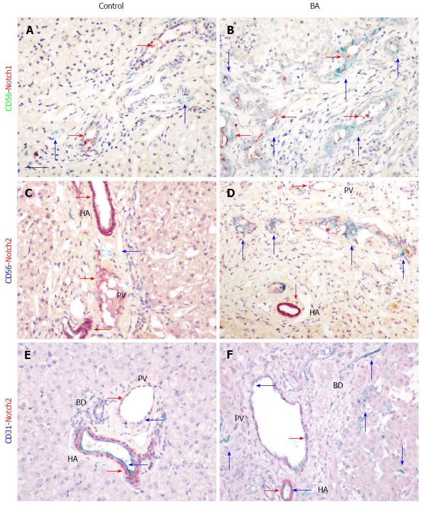
Figure 5 Expression of Notch signaling components in biliary atresia.
Co-localization of CD56-positive cells with Notch1 (A and B) and Notch2 (C and D) is illustrated by immunohistochemical double staining with mouse anti-human CD56 and rabbit anti-human Notch1 and Notch2 using the Polink DS-MR-Hu C1 kit. Positive signals are indicated by arrows, with corresponding colors in the side bars. Scale bar represents 50 μm. BD: Bile duct; HA: Hepatic artery; PV: Portal vein.
- Citation: Zhang RZ, Yu JK, Peng J, Wang FH, Liu HY, Lui VC, Nicholls JM, Tam PK, Lamb JR, Chen Y, Xia HM. Role of CD56-expressing immature biliary epithelial cells in biliary atresia. World J Gastroenterol 2016; 22(8): 2545-2557
- URL: https://www.wjgnet.com/1007-9327/full/v22/i8/2545.htm
- DOI: https://dx.doi.org/10.3748/wjg.v22.i8.2545


