Copyright
©The Author(s) 2016.
World J Gastroenterol. Feb 28, 2016; 22(8): 2545-2557
Published online Feb 28, 2016. doi: 10.3748/wjg.v22.i8.2545
Published online Feb 28, 2016. doi: 10.3748/wjg.v22.i8.2545
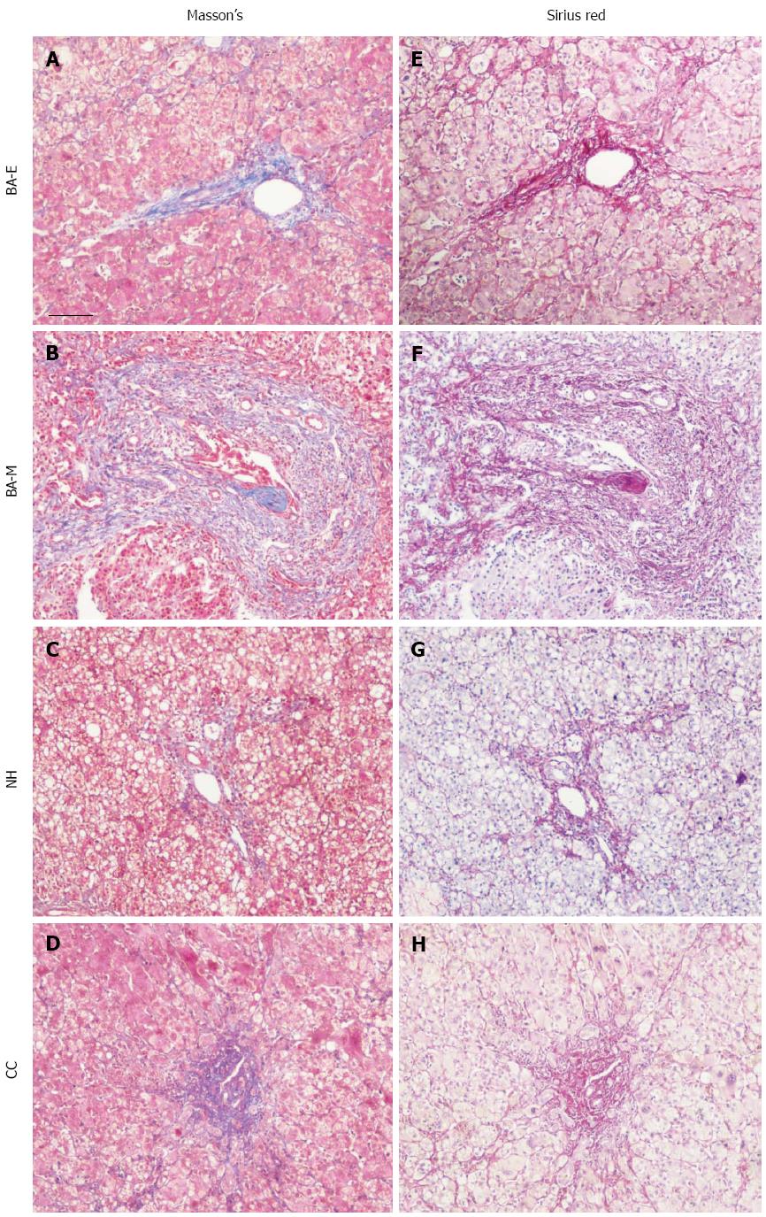
Figure 1 Evaluation of tissue fibrosis in biliary atresia, choledochal cyst, and neonatal hepatitis patients.
Liver tissues from patients with BA, CC, and NH were collected and adjacent sections were cut. Tissue fibrosis was evaluated with Masson’s Trichrome Stain, in which the collagen was detected by blue color. Depending on the stage of fibrosis, sections from patients with BA were further separated into two groups: early stage BA (BA-E), with no or mild collagen deposition, and middle or late stage BA (BA-ML), with dense collagen deposition. The adjacent tissue sections were further stained with Sirius Red to validate the results, with collagen detected by pink color. Sections were examined with a Nikon light microscope. Scale bar shown represents 50 μm. NH: Neonatal hepatitis; CC: Choledochal cyst; BA: Biliary atresia.
- Citation: Zhang RZ, Yu JK, Peng J, Wang FH, Liu HY, Lui VC, Nicholls JM, Tam PK, Lamb JR, Chen Y, Xia HM. Role of CD56-expressing immature biliary epithelial cells in biliary atresia. World J Gastroenterol 2016; 22(8): 2545-2557
- URL: https://www.wjgnet.com/1007-9327/full/v22/i8/2545.htm
- DOI: https://dx.doi.org/10.3748/wjg.v22.i8.2545


