Copyright
©The Author(s) 2016.
World J Gastroenterol. Feb 21, 2016; 22(7): 2398-2402
Published online Feb 21, 2016. doi: 10.3748/wjg.v22.i7.2398
Published online Feb 21, 2016. doi: 10.3748/wjg.v22.i7.2398
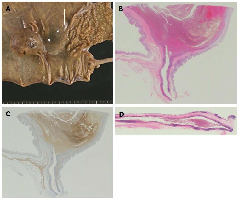
Figure 5 Pathological analysis.
A: A macroscopic view of the resected specimen. A round mass 4 cm in diameter is observed in the ascending colon. A submucosal tubular bulge measuring 5 cm in length extends from the round mass to the ileocecal valve (arrows); B and C: Magnified sectional views of the cystic lesion (loupe images; B: hematoxylin and eosin; C: desmin). The cystic lesion is lined by normal colonic mucosa and possesses its own muscular layer; D: A magnified sectional view of the tubular bulge (loupe image; hematoxylin and eosin). A blind-end tubular structure extends from the cystic lesion toward the proximal side to the ileocecal valve. This structure is lined by normal colonic mucosa and shares the muscular layer with the native ascending colon.
- Citation: Kyo K, Azuma M, Okamoto K, Nishiyama M, Shimamura T, Maema A, Shirakawa M, Nakamura T, Koda K, Yokoyama H. Laparoscopic resection of adult colon duplication causing intussusception. World J Gastroenterol 2016; 22(7): 2398-2402
- URL: https://www.wjgnet.com/1007-9327/full/v22/i7/2398.htm
- DOI: https://dx.doi.org/10.3748/wjg.v22.i7.2398


