Copyright
©The Author(s) 2016.
World J Gastroenterol. Feb 21, 2016; 22(7): 2326-2335
Published online Feb 21, 2016. doi: 10.3748/wjg.v22.i7.2326
Published online Feb 21, 2016. doi: 10.3748/wjg.v22.i7.2326
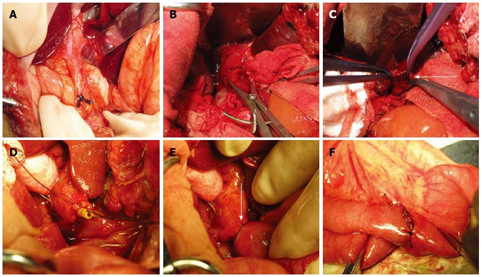
Figure 3 Surgical procedure.
Images illustrating the surgical procedure with a magnet pair. A: The ligation of the distal end of common bile duct near the duodenum. B: Obvious dilatation of the common bile duct can be observed 10 d after ligation; C: Opened common bile duct before placing the biliary part magnet (arrow); D: The biliary part magnet was fixed to the stump of the common bile duct by a purse string; E: The choledochojejunostomy was constructed with the magnet pair (arrow); F: Suture enteroenteric anastomosis between the proximal end of the jejunum and the distal 50 cm of the Roux-en-Y limb.
- Citation: Xue F, Guo HC, Li JP, Lu JW, Wang HH, Ma F, Liu YX, Lv Y. Choledochojejunostomy with an innovative magnetic compressive anastomosis: How to determine optimal pressure? World J Gastroenterol 2016; 22(7): 2326-2335
- URL: https://www.wjgnet.com/1007-9327/full/v22/i7/2326.htm
- DOI: https://dx.doi.org/10.3748/wjg.v22.i7.2326


