Copyright
©The Author(s) 2016.
World J Gastroenterol. Feb 21, 2016; 22(7): 2304-2313
Published online Feb 21, 2016. doi: 10.3748/wjg.v22.i7.2304
Published online Feb 21, 2016. doi: 10.3748/wjg.v22.i7.2304
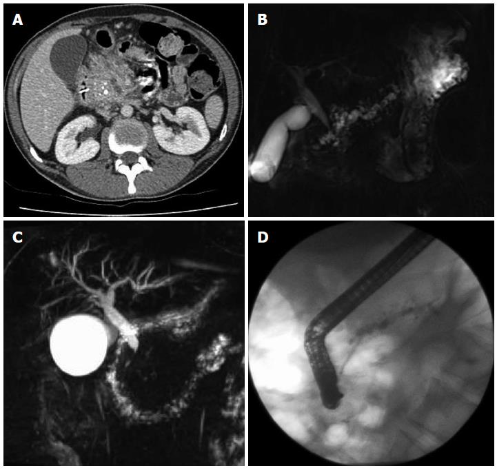
Figure 1 Computed tomography demonstrating enlarged head of pancreas with coarse calcification and a dilated main pancreatic duct (A), magnetic resonance cholangiopancreatography showing a tortuous, dilated pancreatic duct (B), inflammatory stricture of the distal common bile duct (C), endoscopic retrograde cholangiopancreatography showing a stent placed in a dilated pancreatic duct (D).
- Citation: Duggan SN, Ní Chonchubhair HM, Lawal O, O’Connor DB, Conlon KC. Chronic pancreatitis: A diagnostic dilemma. World J Gastroenterol 2016; 22(7): 2304-2313
- URL: https://www.wjgnet.com/1007-9327/full/v22/i7/2304.htm
- DOI: https://dx.doi.org/10.3748/wjg.v22.i7.2304


