Copyright
©The Author(s) 2016.
World J Gastroenterol. Feb 21, 2016; 22(7): 2256-2270
Published online Feb 21, 2016. doi: 10.3748/wjg.v22.i7.2256
Published online Feb 21, 2016. doi: 10.3748/wjg.v22.i7.2256
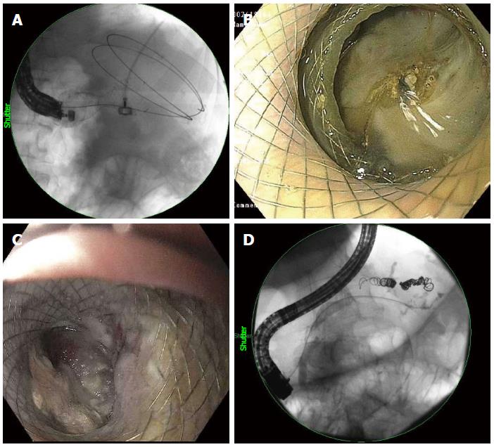
Figure 6 Fluoroscopic visualization of an esophageal fully-covered self-expanding metal stents deployed into walled-off necrosis (A), a pancreatic duct leak (D), endoscopic visualization of a lumen-apposing metal stent deployed into walled-off necrosis (B, C).
- Citation: Tyberg A, Karia K, Gabr M, Desai A, Doshi R, Gaidhane M, Sharaiha RZ, Kahaleh M. Management of pancreatic fluid collections: A comprehensive review of the literature. World J Gastroenterol 2016; 22(7): 2256-2270
- URL: https://www.wjgnet.com/1007-9327/full/v22/i7/2256.htm
- DOI: https://dx.doi.org/10.3748/wjg.v22.i7.2256


