Copyright
©The Author(s) 2016.
World J Gastroenterol. Feb 14, 2016; 22(6): 2153-2158
Published online Feb 14, 2016. doi: 10.3748/wjg.v22.i6.2153
Published online Feb 14, 2016. doi: 10.3748/wjg.v22.i6.2153
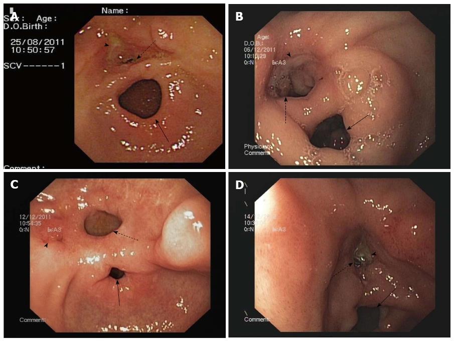
Figure 1 Double pylorus observed.
A: A 41-year-old man undergoing endoscopy due to epigastric pain. A yellow-based irregular ulcer (arrowhead) is present in the antrum of the lesser curve over the accessory pylorus (dotted arrow). The solid arrow indicates the true pylorus; B: A 61-year-old man with osteoarticular degenerative disease who underwent endoscopy due to melena. A white-based ulcer (arrowhead) with edematous margins within the accessory pylorus (dotted arrow) on the lesser curve of the peri-pyloric region is present. The other opening is the normal pylorus (solid arrow); C: A 58-year-old man with headache who underwent endoscopy due to coffee-ground vomitus and melena. A white-based deep ulcer (arrowhead) is present in the anterior wall of the gastric antrum on the left side of the accessory pylorus (dotted arrow). The solid arrow indicates the true pylorus. D: A 62-year-old woman with gout who underwent endoscopy due to abdominal pain. A white-based ulcer (arrowhead) within the accessory pylorus (dotted arrow) is visible. The other opening is the true pylorus (solid arrow). Severe erythematous gastritis of the antrum is also present.
- Citation: Lei JJ, Zhou L, Liu Q, Xu CF. Acquired double pylorus: Clinical and endoscopic characteristics and four-year follow-up observations. World J Gastroenterol 2016; 22(6): 2153-2158
- URL: https://www.wjgnet.com/1007-9327/full/v22/i6/2153.htm
- DOI: https://dx.doi.org/10.3748/wjg.v22.i6.2153


