Copyright
©The Author(s) 2016.
World J Gastroenterol. Feb 14, 2016; 22(6): 2092-2103
Published online Feb 14, 2016. doi: 10.3748/wjg.v22.i6.2092
Published online Feb 14, 2016. doi: 10.3748/wjg.v22.i6.2092
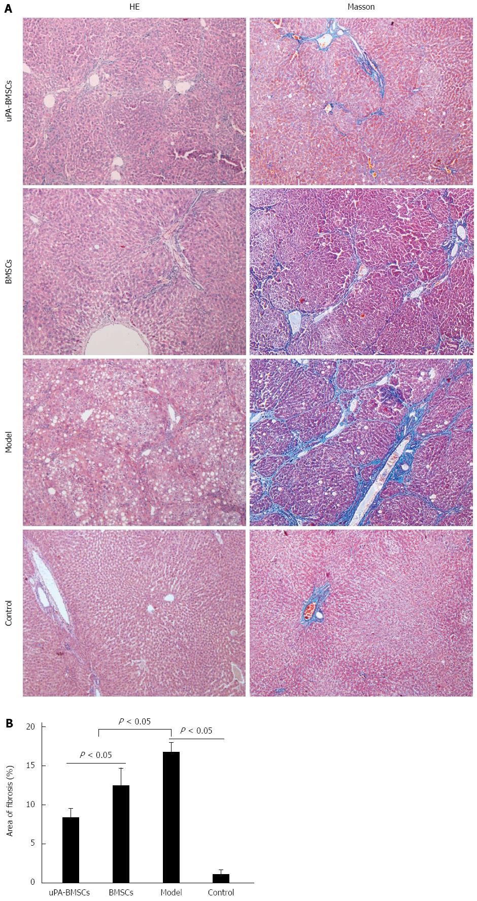
Figure 3 Histopathological change of liver tissue in different groups.
A: HE and Masson staining was used to detect structural changes in liver tissues; B: Quantitative analyzes of liver fibrosis were performed using Masson stained sections. Five random views from each sample in each group were analyzed. BMSCs: Bone marrow-derived mesenchymal stem cells; uPA: Urokinase plasminogen activator.
- Citation: Ma ZG, Lv XD, Zhan LL, Chen L, Zou QY, Xiang JQ, Qin JL, Zhang WW, Zeng ZJ, Jin H, Jiang HX, Lv XP. Human urokinase-type plasminogen activator gene-modified bone marrow-derived mesenchymal stem cells attenuate liver fibrosis in rats by down-regulating the Wnt signaling pathway. World J Gastroenterol 2016; 22(6): 2092-2103
- URL: https://www.wjgnet.com/1007-9327/full/v22/i6/2092.htm
- DOI: https://dx.doi.org/10.3748/wjg.v22.i6.2092


