Copyright
©The Author(s) 2016.
World J Gastroenterol. Dec 28, 2016; 22(48): 10482-10501
Published online Dec 28, 2016. doi: 10.3748/wjg.v22.i48.10482
Published online Dec 28, 2016. doi: 10.3748/wjg.v22.i48.10482
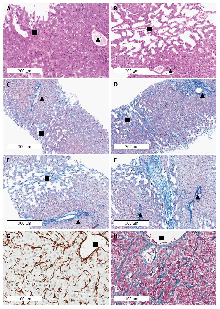
Figure 6 Cardiac cirrhosis as example of a pressure-associated cirrhosis in the absence of notable inflammation.
Patients with congestive heart failure may even die from complications of cirrhosis such as variceal bleeding or liver cancer. Note the absence of inflammation in areas with sinusoidal dilation and congestion in long-lasting congestion characterized by marked sinusoidal dilation and atrophy of liver cell plates. Early (A) and advanced (B) stages of congestive heart failure stained by hematoxyline and eosin. Portal tracts and centrilobular areas are marked with black triangles and squares, respectively. Inflammation is also not a feature in areas with sinusoidal dilation and congestion in intermediate and central portions of the lobules. C: Chromotrope aniline blue stain (fibrosis) of an early stage of congestive hepatopathy in a case with congestive heart failure. The portal tract and its structures is regular whereas in central and intermediate portions of the hepatic lobulus mild sinusoidal dilation, slight atrophy of liver cell plates and minimal perisinusoidal fibrosis are seen. D: If venous outlaw obstruction persists perisinusoidal fibrosis and atrophy of liver cell plates in centrilobular areas become more pronounced, (E) which is then followed by loss of liver cell plates and centrilobular fibrosis extending towards neighbouring central veins (F) finally resulting in fibrous septa (marked by arrow heads). Notably, portal-central relations are mostly preserved. Stain for αSMA (G) and fibrosis (Masson trichrome) (H) indicating septal and perisinusoidal fibrosis from a liver biopsy of a 31 years-old male patient with Fontan circulation. The images show diffuse activation of hepatic stellate cells in the absence of any inflammation. Fontan intervention was performed early around birth because of an unilateral ventricle. HVPG was 1 mmHg, LS was 19 kPa. (Images A-F: Courtesy of Dr. C. Lackner, University of Graz; images G-H: Courtesy of Dr. P. Bedossa, Hôpital Beaujon, Université Paris Diderot). HVPG: Hepatic venous pressure gradient; LS: Liver stiffness.
- Citation: Mueller S. Does pressure cause liver cirrhosis? The sinusoidal pressure hypothesis. World J Gastroenterol 2016; 22(48): 10482-10501
- URL: https://www.wjgnet.com/1007-9327/full/v22/i48/10482.htm
- DOI: https://dx.doi.org/10.3748/wjg.v22.i48.10482


