Copyright
©The Author(s) 2016.
World J Gastroenterol. Dec 21, 2016; 22(47): 10353-10363
Published online Dec 21, 2016. doi: 10.3748/wjg.v22.i47.10353
Published online Dec 21, 2016. doi: 10.3748/wjg.v22.i47.10353
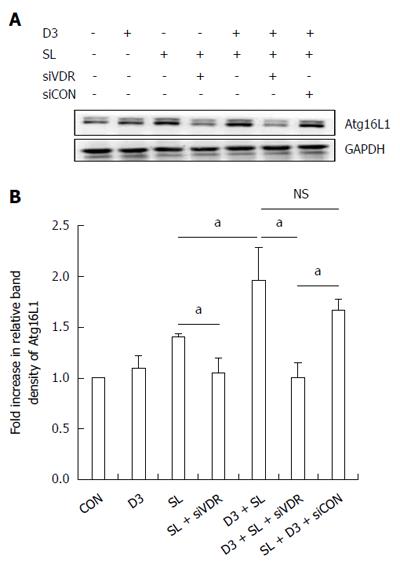
Figure 4 Effect of 1,25D3 on the membranous Atg16L1 protein expression in Salmonella-infected Caco-2 cells via vitamin D receptor.
Caco-2 cells were transfected with control siRNA and vitamin D receptor (VDR) siRNA (siCON: non-target control siRNA; siVDR: siRNA to VDR) for 48 h. The transfected cells were left uninfected (CON) or infected by wild-type S. typhimurium strain SL1344 (SL) in the absence or presence of 1,25D3 (D3). Immunoblots were performed on membrane lysates with antibody to detect Atg16L1 protein expression, and E-cadherin for normalization of membrane proteins. Representative immunoblots (A) and densitometric quantification of immunoreactive bands (B) are shown. The relative band intensities of Atg16L1 in Caco-2 cells were quantified as fold-increases compared with the control cells. Each value represents the mean ± SEM of three independent experiments. aP < 0.05.
- Citation: Huang FC. Vitamin D differentially regulates Salmonella-induced intestine epithelial autophagy and interleukin-1β expression. World J Gastroenterol 2016; 22(47): 10353-10363
- URL: https://www.wjgnet.com/1007-9327/full/v22/i47/10353.htm
- DOI: https://dx.doi.org/10.3748/wjg.v22.i47.10353


