Copyright
©The Author(s) 2016.
World J Gastroenterol. Dec 21, 2016; 22(47): 10287-10303
Published online Dec 21, 2016. doi: 10.3748/wjg.v22.i47.10287
Published online Dec 21, 2016. doi: 10.3748/wjg.v22.i47.10287
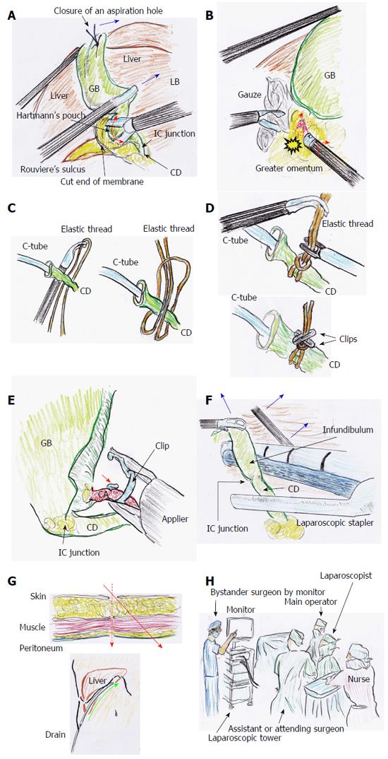
Figure 5 Tips and pitfalls of laparoscopic cholecystectomy.
A: After GB decompression by aspiration, the GB neck and/or Hartmann’s pouch can be pulled from the dorsal space. Hence, dissection can be performed as close to the GB as possible (red arrow) under adequate retractions (blue arrow); B: The rubbing of a bleeding vessel or oozing tissue (dotted arrow) by a button-shaped electrode with suction with a soft-coagulation system is a key technique for reliable hemostasis. During this hemostasis, subtle rotation of the electrode is important (red arrow); C: An elastic thread is never ligated directly; D: Clips are positioned to establish angular separation; E: A clip should be applied with the tip extending beyond the duct or vessels (red arrow); F: If the CD is too thick, loop ligation or a laparoscopic stapler can be chosen, under adequate retractions (blue arrows); G: Laparoscopic port penetrates abdominal wall at right angle (dotted arrow). A drain pathway through the abdominal wall is remade from the same skin incision (red arrow), to make the best drain placement (green arrow); H: A detached observer may be an actual solution for prevention of misidentification during LC. CD: Cystic duct; GB: Gallbladder; IC: Infundibulum-cystic duct; LC: Laparoscopic cholecystectomy.
- Citation: Hori T, Oike F, Furuyama H, Machimoto T, Kadokawa Y, Hata T, Kato S, Yasukawa D, Aisu Y, Sasaki M, Kimura Y, Takamatsu Y, Naito M, Nakauchi M, Tanaka T, Gunji D, Nakamura K, Sato K, Mizuno M, Iida T, Yagi S, Uemoto S, Yoshimura T. Protocol for laparoscopic cholecystectomy: Is it rocket science? World J Gastroenterol 2016; 22(47): 10287-10303
- URL: https://www.wjgnet.com/1007-9327/full/v22/i47/10287.htm
- DOI: https://dx.doi.org/10.3748/wjg.v22.i47.10287


